central axillary lymph nodes
Lymph nodes on mammogram of the right breast | Image | Radiopaedia.org. 11 Pics about Lymph nodes on mammogram of the right breast | Image | Radiopaedia.org : Regional Lymph Nodes | SEER Training, Breast Cancer Lymph Node Surgery | Manhattan, NY and also Regional Lymph Nodes | SEER Training.
Lymph Nodes On Mammogram Of The Right Breast | Image | Radiopaedia.org
 radiopaedia.org
nodes lymph mammogram breast radiopaedia right version
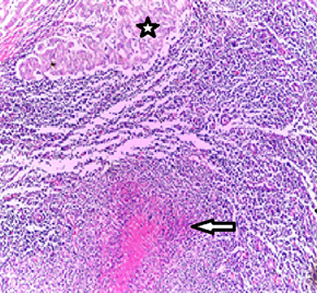
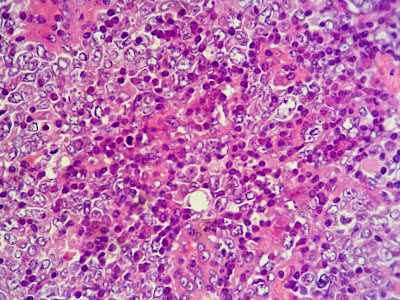
Anatomo-Clinical Case: Coexistence Of Tuberculosis With Axillary Lymph
 www.scirp.org
tuberculosis lymph carcinoma infiltrating eosin proliferation carcinomatous magnification
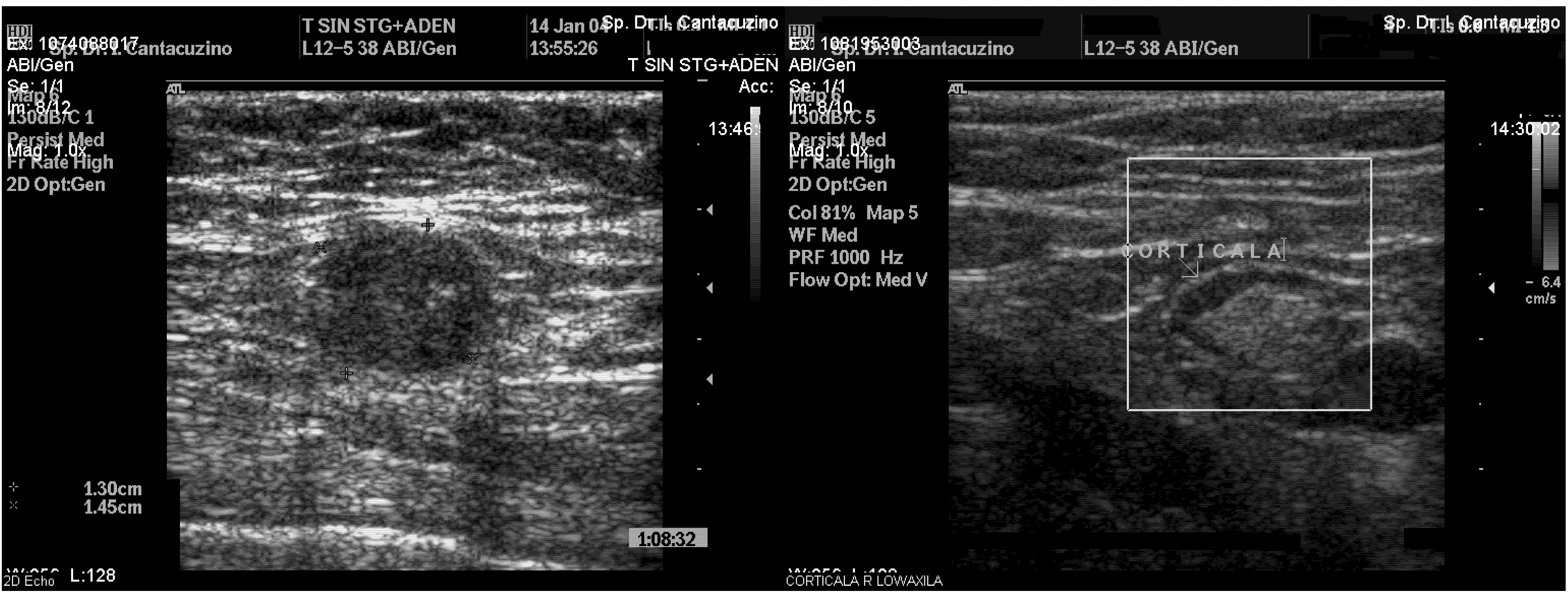
Ultrasound Axillary Imaging | IntechOpen
 www.intechopen.com
ultrasound axillary imaging intechopen figure
A Gallery Of High-Resolution, Ultrasound, Color Doppler & 3D Images
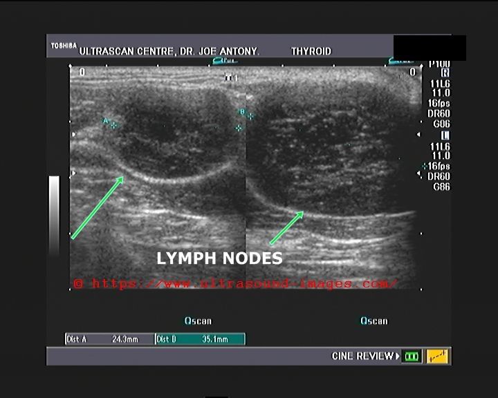 www.ultrasound-images.com
lymphoma lymph hodgkins
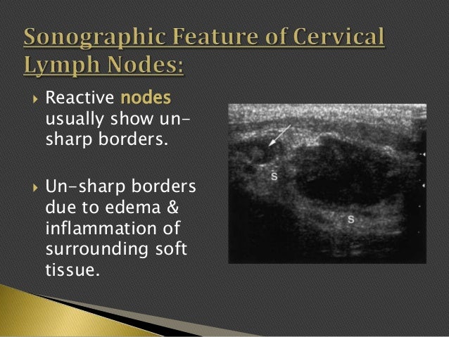
Imaging Of Enlarged Lymph Node
 www.slideshare.net
lymph reactive
High-resolution Ultrasonographic Features Of Axillary Lymph Node
 www.thebreastonline.com
lymph node breast axillary cancer metastasis features fig nodes ultrasound ultrasonographic patients resolution normal examination
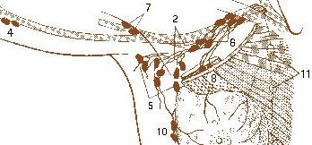
Breast Cancer Lymph Node Surgery | Manhattan, NY
 www.laparoscopicsurgeons.com
lymph axillary node surgery nodes level surgical minor breast pectoralis label cancer depths lay different three
HISTOPATHOLOGY: Axillary Lymph Node (15)
 histopathologyview.blogspot.com
axillary lymph histopathology
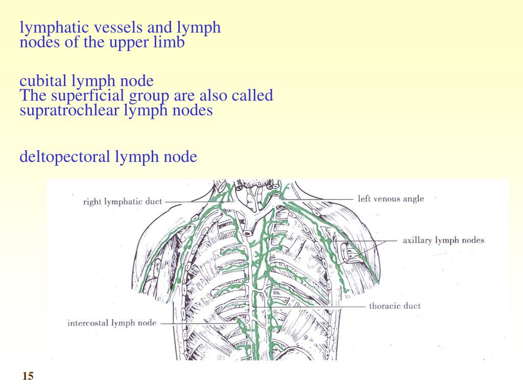
PPT - Main Collecting Lymphatic Channels Lymphatic Drainage Of The Head
 www.slideserve.com
lymph lymphatic head node ppt drainage collecting channels neck main nodes pectoral subscapular cubital lateral supratrochlear powerpoint presentation
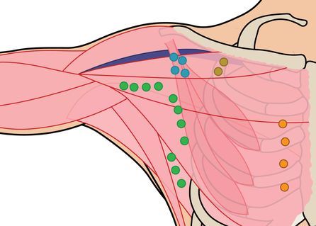
Regional Lymph Nodes | SEER Training
 www.training.seer.cancer.gov
lymph nodes breast axillary parasternal cancer subscapular node regional training cubital anatomy apical lymphatic intramammary seer rotter wikipedia illustration mammary
Color Doppler Ultrasound Image Of Benign Lymph Node Showing
 www.researchgate.net
lymph ultrasound doppler benign vascularity hilar
Anatomo-clinical case: coexistence of tuberculosis with axillary lymph. Ultrasound axillary imaging intechopen figure. Imaging of enlarged lymph node