chest and stomach anatomy
Hypertrophy Left Ventricle with Thickened Wall - Chest Case Studies. 9 Images about Hypertrophy Left Ventricle with Thickened Wall - Chest Case Studies : Innervation of the heart: Sympathetic and parasympathetic | Kenhub, VET Talks - Normal Radiographic Anatomy of the Canine Thorax - YouTube and also Pulmonary Edema in Patient with Interstitial Lung Disease - Chest Case.
Hypertrophy Left Ventricle With Thickened Wall - Chest Case Studies
 www.ctisus.com
ventricle ctisus hypertrophy
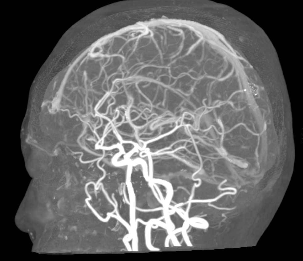
CTA Of Circle Of Willis - Neuro Case Studies - CTisus CT Scanning
 www.ctisus.com
cta ct brain ctisus willis circle neuro scan studies case diagnosis scanning
Asplenia Syndrome | Image | Radiopaedia.org
 radiopaedia.org
asplenia radiopaedia
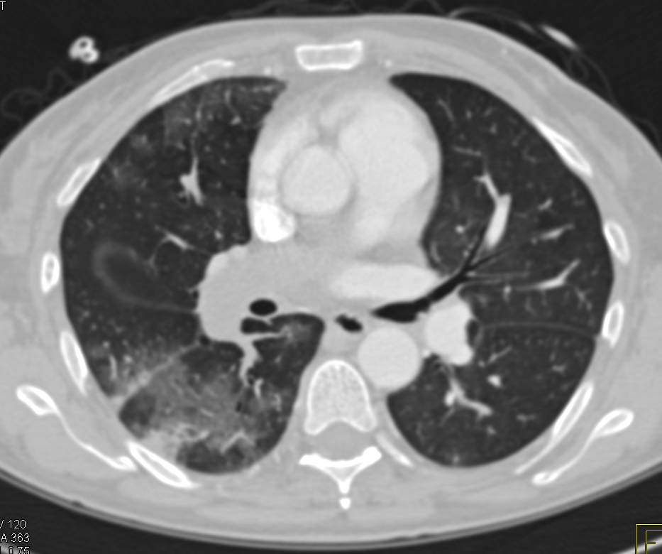
Large Pulmonary Embolism With Lung Infarct And Splenic Infarct - Chest
 www.ctisus.com
ct pulmonary chest embolism infarct ctisus scan lung diagnosis studies case
Pulmonary Edema In Patient With Interstitial Lung Disease - Chest Case
 ctisus.com
interstitial edema ctisus pulmonary
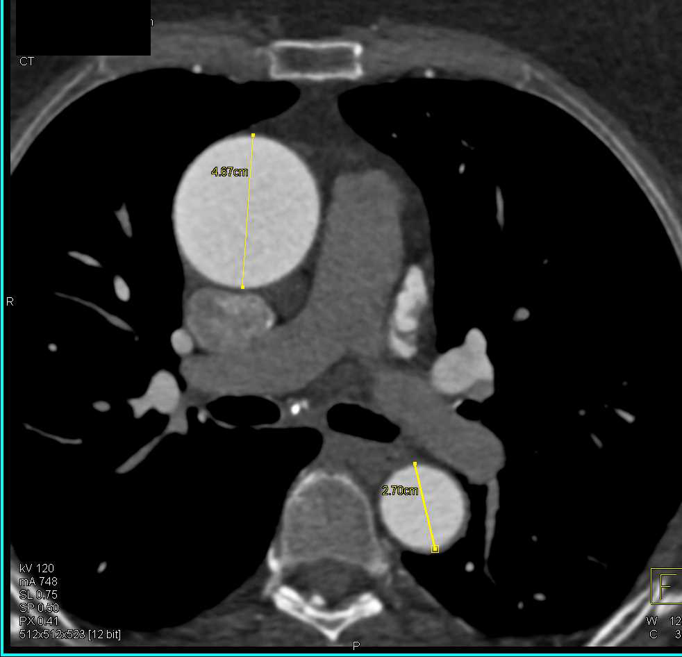
Dilated Ascending Aorta - Chest Case Studies - CTisus CT Scanning
 www.ctisus.com
aorta ascending ctisus aortic leaflets cardiac dilated
Innervation Of The Heart: Sympathetic And Parasympathetic | Kenhub
heart nerve phrenic anatomy innervation nervus left thorax nerves kenhub sympathetic plexus sinister parasympathetic thoracic path neck through supply brachial
Focal Nodular Hyperplasia | Image | Radiopaedia.org
 radiopaedia.org
nodular hyperplasia focal liver ultrasound normal abdominal gallbladder abnormal dynamic range radiopaedia case steatosis version appeared usual settings under finding
VET Talks - Normal Radiographic Anatomy Of The Canine Thorax - YouTube
 www.youtube.com
canine normal anatomy thorax radiographic vet
Cta of circle of willis. Dilated ascending aorta. Ct pulmonary chest embolism infarct ctisus scan lung diagnosis studies case