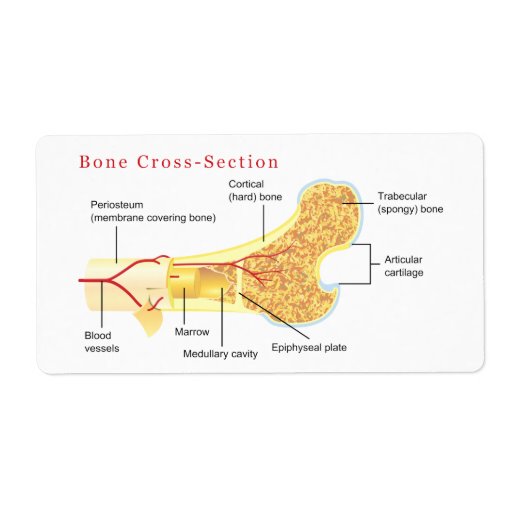
cross section of bone labeled
Bone and Bone Formation | histology. 11 Pics about Bone and Bone Formation | histology : Bone Cross Section Diagram Labels | Zazzle, Bone Cross Section Labeled - Cross Section Of A Bone Cross Section Of and also Chapter 7, Page 6 - HistologyOLM.
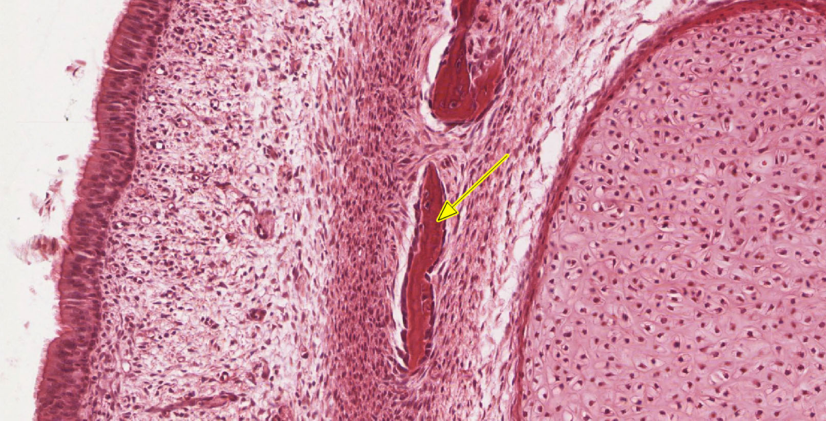
Bone And Bone Formation | Histology
 histology.medicine.umich.edu
bone formation histology ossification microscope endochondral remodeling intramembranous under cartilage center calcified surface
The Canine Head And Skull (CT): Atlas Of Veterinary Clinical And
 www.clinicalelearning.com
canine paranasal orbital labeled ethmoid cranium basisphenoid
Bone Cross Section Diagram Labels | Zazzle
 www.zazzle.com
bone section cross diagram labels label shipping zazzle
Bone Gallery
 medcell.med.yale.edu
muscle skeletal section cross labeled diagram histology cell bone
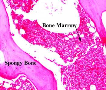
10 Tissues Lab
 bio1151.nicerweb.com
bone tissue lab tissues marrow adipose spongy increasing nicerweb locked growth interstitial
Human Bone Cross Section Diagram Of Femur Showing Osteon Veins Marrow
 www.gettyimages.com
bone section cross femur diagram human osteon marrow showing illustration veins
Digestive | NP Histology
 sites.newpaltz.edu
histology esophagus 4x section cross viewed objective anatomy digestive
Bone Cross Section Labeled - Cross Section Of A Bone Cross Section Of
 doditdewantara.blogspot.com
tissue
Chapter 7, Page 5 - HistologyOLM
 www.stevegallik.org
muscle skeletal section cross histology tissue normal microscopic histologyolm study stevegallik chapter preparation
Chapter 7, Page 6 - HistologyOLM
 www.stevegallik.org
neuromuscular junction muscle nerve skeletal motor synapse tissue microscopic where neuron junctions microscopy lab meets identify 3d end fiber structure
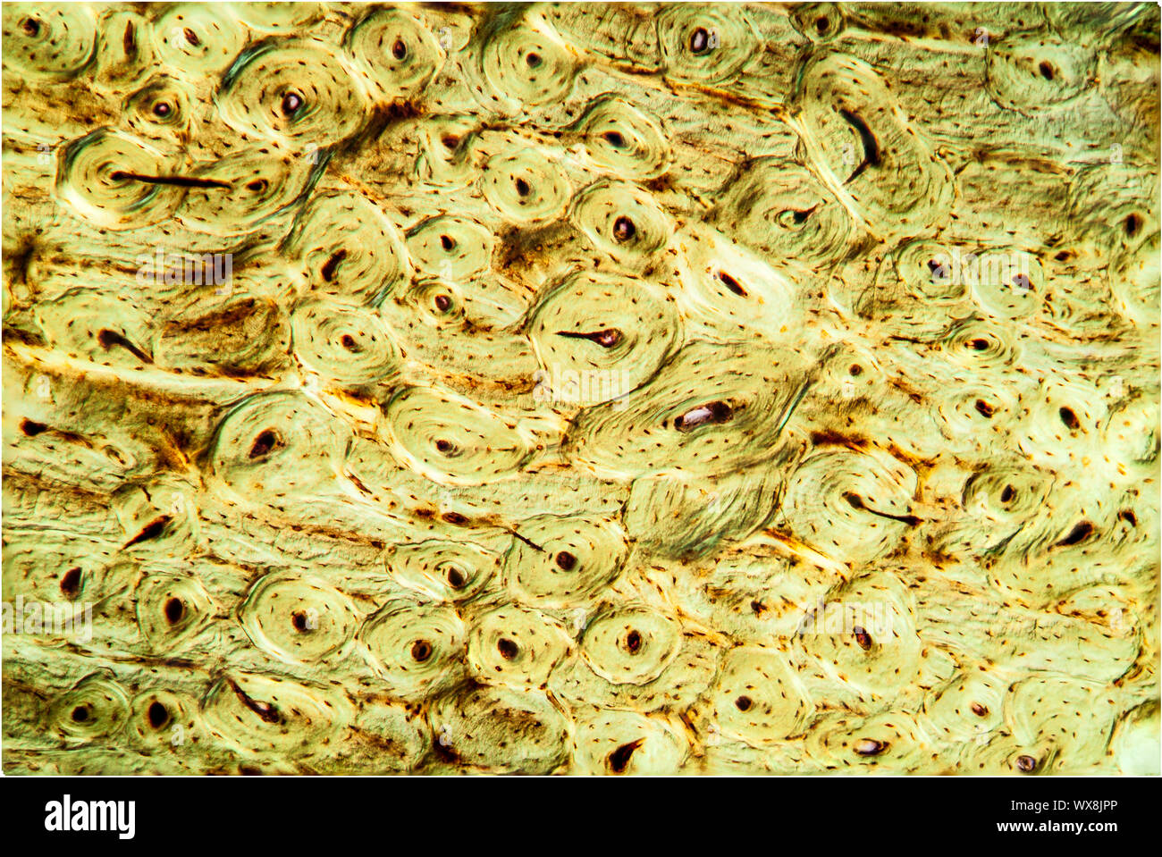
Chapter 6, Page 5 - HistologyOLM
 www.stevegallik.org
bone compact decalcified low magnification human preparation histologyolm microscopic fig stevegallik
Bone formation histology ossification microscope endochondral remodeling intramembranous under cartilage center calcified surface. Muscle skeletal section cross histology tissue normal microscopic histologyolm study stevegallik chapter preparation. Bone section cross femur diagram human osteon marrow showing illustration veins