inferior mesenteric artery location
PPT - Chapter 21 PowerPoint Presentation, free download - ID:762038. 10 Pictures about PPT - Chapter 21 PowerPoint Presentation, free download - ID:762038 : Superior Mesenteric Artery (SMA) - Stepwards, Vascular Anatomy of the Pelvis | Radiology Key and also Pin by Ellis Joan on Abd Ultrasound 100 Mod 2 | Ultrasound sonography.
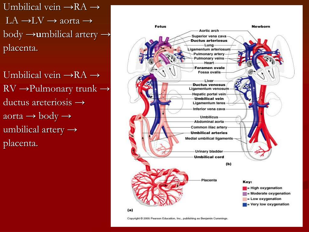
PPT - Chapter 21 PowerPoint Presentation, Free Download - ID:762038
 www.slideserve.com
umbilical vein arteries powerpoint artery placenta chapter ppt presentation aorta veins blood
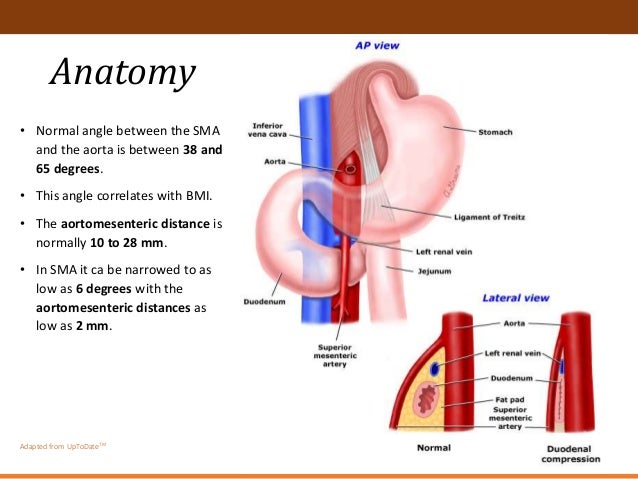
Superior Mesenteric Artery Syndrome
 www.slideshare.net
syndrome artery mesenteric
View Image
 www.eusjournal.com
vein mesenteric superior splenic ultrasound doppler artery inferior portal pancreas imaging trunk left right joins tributaries close linear endoscopic junction
Arteries & Veins - Atlas Of Anatomy
 doctorlib.info
anatomy artery mesenteric superior celiac trunk arteries atlas fig anterior veins doctorlib medical info
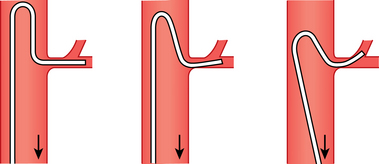
Mesenteric Arteries | Radiology Key
 radiologykey.com
mesenteric arteries catheter sidewinder
Pin By Ellis Joan On Abd Ultrasound 100 Mod 2 | Ultrasound Sonography
 www.pinterest.ie
aorta sagittal liver gastroesophageal sonography artery mesenteric uonbi
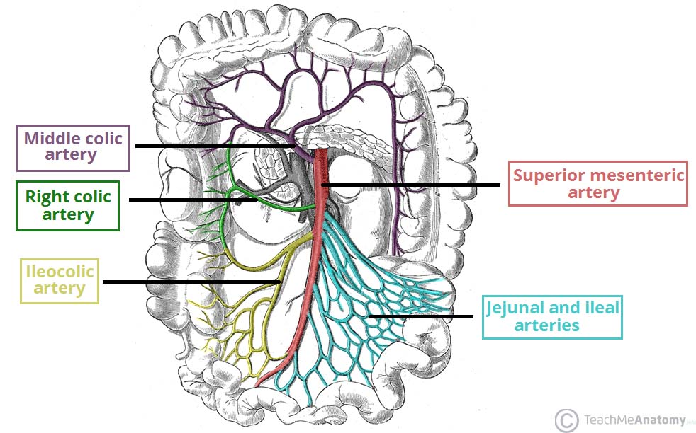
Superior Mesenteric Artery (SMA) - Stepwards
 www.stepwards.com
mesenteric superior artery branches sma inferior anatomy supply blood vessels its arteries midgut aorta arterial pancreaticoduodenal ischaemia stepwards vasculature vascular
Superior Mesenteric Artery - Wikidoc
 www.wikidoc.org
mesenteric superior artery sma branches vein vessel wikidoc frontal beside its
Photographs Of The Vessels Of The Fetal Pig
 blog.valdosta.edu
pig fetal vein artery veins vessels anterior aorta exposed mammary left internal
Vascular Anatomy Of The Pelvis | Radiology Key
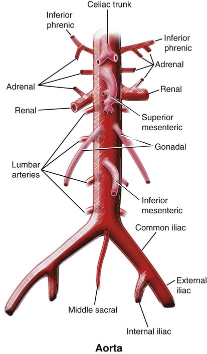 radiologykey.com
iliac abdominal arteries anatomy common vascular pelvis aorta artery branches bifurcation internal external abdomen left into major renal gonadal inferior
Vascular anatomy of the pelvis. Pig fetal vein artery veins vessels anterior aorta exposed mammary left internal. Photographs of the vessels of the fetal pig